
From PDB to Production: How to Prep Protein Structures for 3D Animation
We are frequently asked to create animations that depict complex interactions between molecules. As medical illustrators, it’s essential to deeply understand the molecular processes we illustrate. Our goal is to convey key scientific information with clarity and precision, making it accessible to our audience, whether they are researchers, clinicians, or patients.
Step 1: Initial Research and Understanding
Our process begins with thorough research to develop a solid understanding of the topic. This involves reviewing current literature surrounding the protein interaction. The protein in question may play a role in disease pathophysiology or serve as a therapeutic target. Understanding this context, along with the client’s brief, allows us to ask the right questions:
Where is the protein located in the body or cell?
Is it part of a larger complex?
Is there a ligand involved?
If so, where are the binding pockets or interaction regions?
These questions shape our research strategy and guide how we visualize the story.
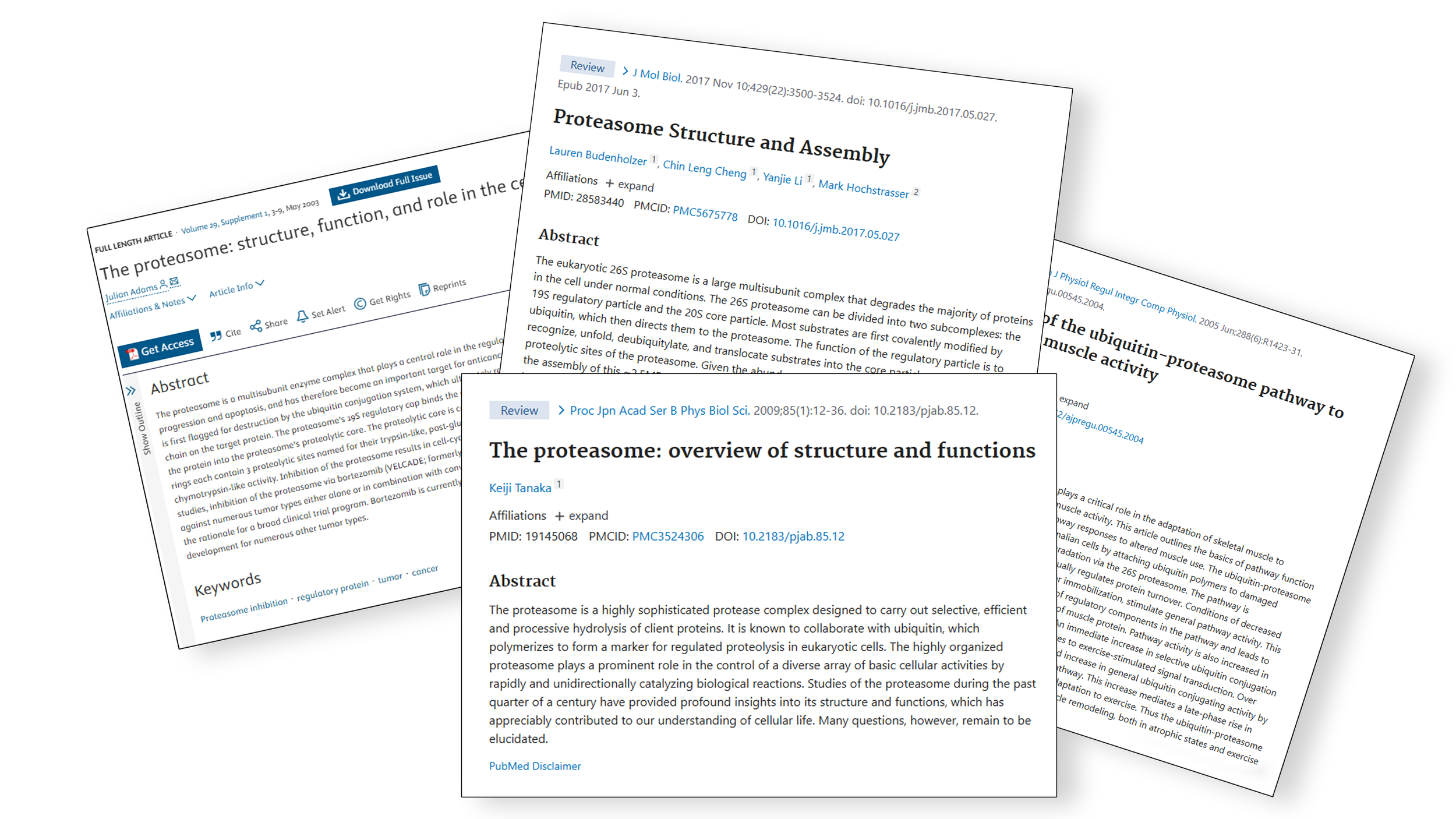
Step 2: Gathering Evidence and Structural Data
Once we’ve defined the key questions, we begin collecting peer-reviewed research papers and relevant data sources. These help inform the scientific basis of our visual storytelling.
We also consult several specialized biological databases to access accurate structural and functional information:
Protein Data Bank (PDB)
A critical resource for 3D structural data of proteins, nucleic acids, and complexes. It's especially valuable for medical illustrators as it allows us to:
Visualize protein folding and conformation
Identify active/binding sites
Explore structural changes due to ligand binding or mutations
UniProt
This is a comprehensive protein database providing:
Sequence and functional data
Expression profiles and cellular location
Information on domains, interactions, and disease relevance
Curated links to literature and other databases
UniProt is helpful when determining if a protein is expressed in a particular organelle or tissue, which can inform the design of broader cellular or anatomical scenes.
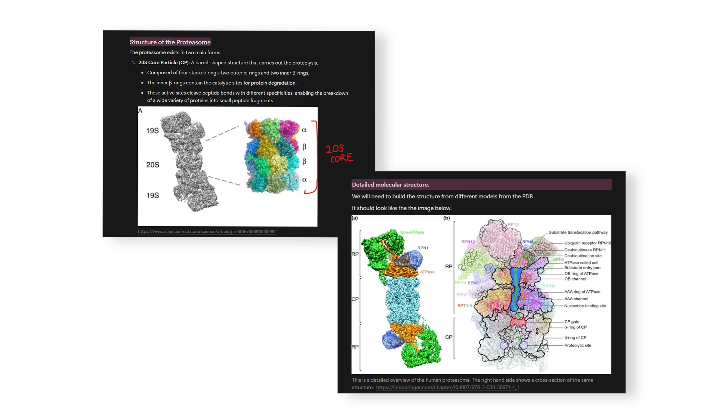
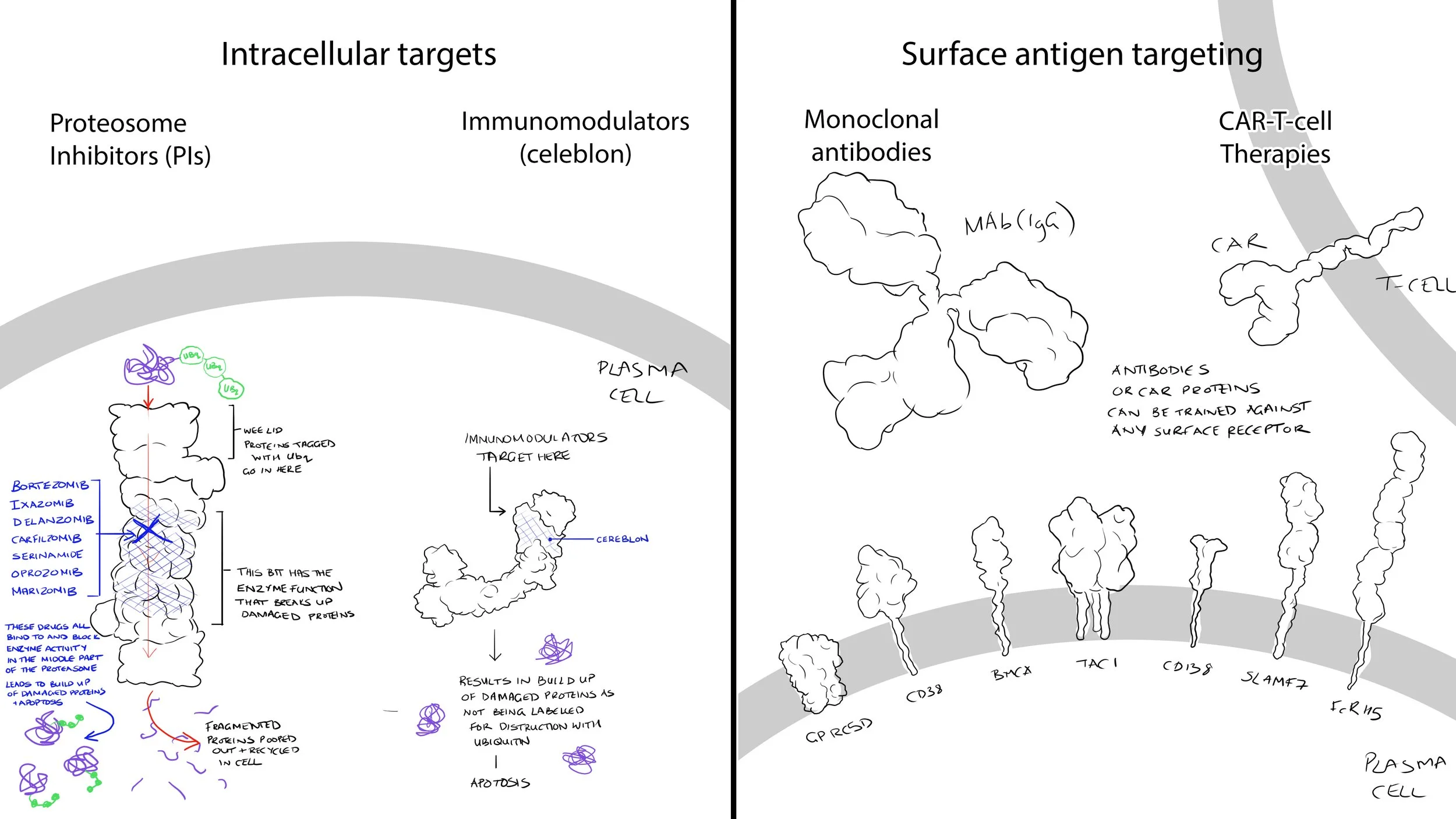
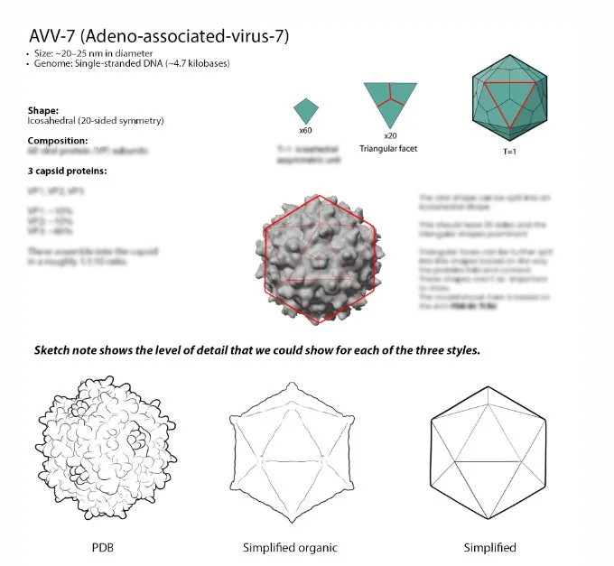
Step 3: Organizing Research into Visual Resources
We distill our findings into internal research cards. These are summarized, visual documents that compile all relevant structural, functional, and contextual information. These cards become part of our internal library and serve as a reference throughout the project.
We often create sketchnotes. These are visual summaries of complex topics to communicate ideas internally and externally. These help unify the team’s vision and ensure scientific accuracy throughout production.
Step 4: Preparing Structures for Animation
1. Download and Edit Protein Models
Use the PDB to download the protein’s structure file (e.g.,
.pdbor.glb)Use Chimera to edit the model, add representations (e.g., ribbons, surfaces), or visualize interactions with ligands or other molecules
2. Import into Cinema 4D
Export the processed model from Chimera and import it into Cinema 4D (C4D)
Clean up the geometry, group structures logically, and label parts clearly
3. Optimize Mesh for Animation
PDB models can be highly detailed, resulting in dense meshes
Use remeshing or volume building tools in C4D to reduce polygon count while retaining shape fidelity
Align and scale molecules to match other scene components based on research findings
Step 5: Storyboarding and Team Communication
We condense all this information into a clear guide for the storyboard artists and animators. This includes:
Key concepts and molecular events to visualize
Scene composition and camera movements
Client branding or visual style guidelines
We often create notes along with our storyboards to help guide our team of animators on subtle movements and styles as well as things to avoid.
Why This Matters
Understanding protein interactions and structural biology is essential in medical animation. Proteins are the functional engines of biology, governing everything from cellular communication to drug action. Accurate visualization grounded in structural data:
Makes complex mechanisms accessible and engaging
Supports scientific storytelling in education and communication
Provides clarity in visualizing disease pathways or therapeutic interventions
We hope you enjoyed this behind-the-scenes look into our process. Stay tuned for more insights on how we continue to blend creativity and science in our projects! 🚀
Written by: Dr Angela Douglass, Lead Researcher and Medical Illustrator at Now Medical Studios